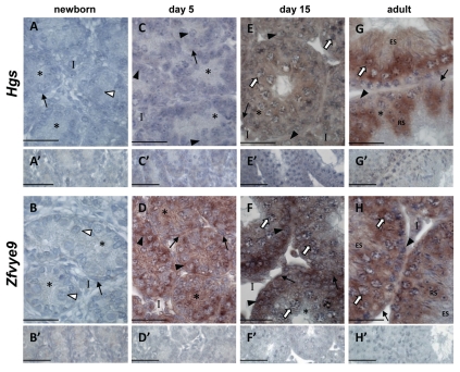
Figure 3.
Hgs and Zfyve9 are differentially expressed in the developing postnatal and adult mouse testis. In situ hybridisation using DIG-labeled riboprobes localizes Hgs and Zfyve9 mRNAs (purple staining) in mouse testis sections at the indicated ages, counterstained with Harris Haematoxylin (blue) to visualize chromatin. In all cases, no signal was detected when sections were incubated with sense probe (A′–H′). White arrowhead: gonocytes; black arrow head: spermatogonia; white arrow: spermatocytes; RS: round spermatids; ES: elongating spermatids; asterisk: Sertoli cell cytoplasm; black arrow: peritubular myoid cells; I: interstitium. Scale bars = 50 µm.

