Fig. 1.
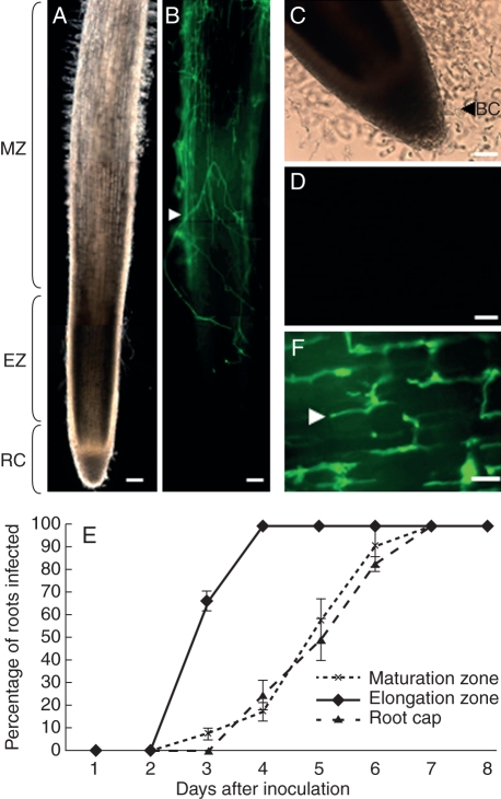
Localization of A. euteiches (RB 84) in the root tip of P. sativum. (A) Bright field image of the primary root tips at 4 d post-inoculation and (B) FITC–WGA-staining showing the presence of mycelia on the root surface. (C) Bright field image showing the release of border cells from the root cap at 4-d post-inoculation. (D) Note the absence of mycelia on the root cap and border cells as revealed by FITC–WGA staining. (E) Time-course progression of A. euteiches within the root tissues in the elongation zone, maturation zone and root caps. The presence of mycelia was detected on root sections using FITC–WGA staining (n = 30 roots) Values are means ± standard deviation from three independent experiments. (F) By 3-d post-inoculation the mycelium was abundant in the root cortex of the elongtion zone. BC, Border cells; EZ, root elongation zone; RC, root cap; MZ, root maturation zone. White arrowheads indicate the presence of mycelia. Scale bars: (A, B) = 100 µm; (C, D) = 50 µm; (F) = 5 µm.

