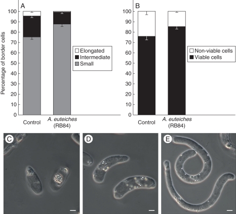
Fig. 7.
Effect of A. euteiches infection on both (A) border cell morphology and (B) viability analysed by flow cytometry. (C–E) Micrographs showing the three types of border cells released by the root tip of P. sativum: small spherical cells (C), intermediate-size cells (D) and elongated cells (E). Scale bars = 5 µm.

