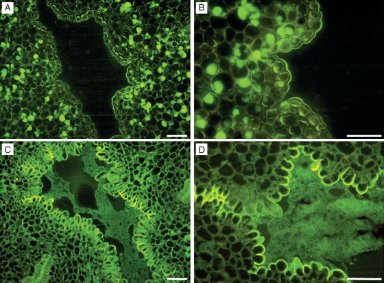
Fig. 3.
Central channel modification during flower maturation in ‘Nova’ mandarin. (A) At anthesis, in the central channel, an empty space can be observed, which is bordered by cells with thick walls (B). Some of the cells in (A) and (B) show stained lipidic contents. (C) Four days after flower-opening, cells bordering the central channel changed their appearance and entered a secretory phase (D) as the cells became papillated. JB-4-embedded transverse sections of the central canal stained with auramine. Scale bars = 50 µm.

