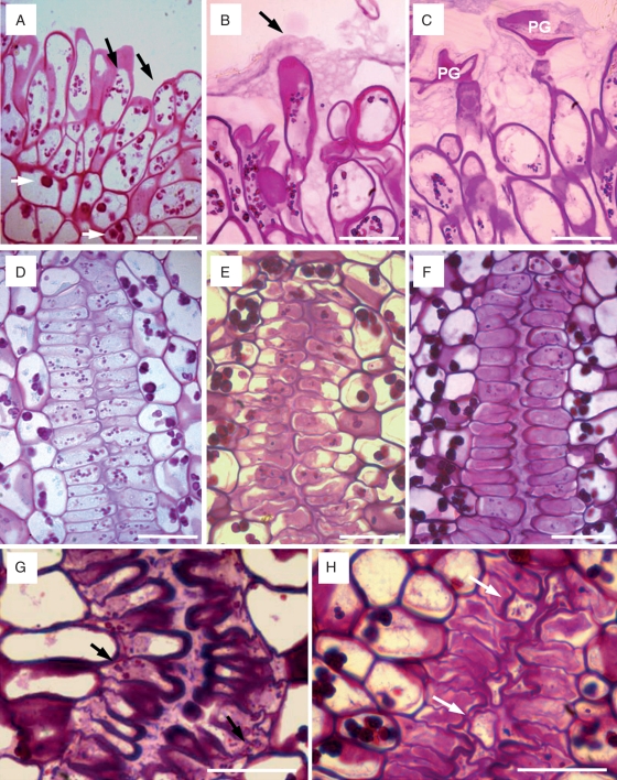
Fig. 4.
Changes in the stigma and style in ‘Nova’ mandarin. (A) Stigmatic papillae at anthesis with small starch grains (black arrows) with a different appearance than starch in parenchymatic cells (white arrows). (B) Stigmatic papillae 2 d after anthesis covered with a stigmatic compact secretion (black arrow). (C) Germinated pollen grain (PG) in degraded stigmatic secretion in a flower 2 d after pollination. (D) Cells bordering the stylar canal with starch at anthesis. (E, F) Progressive degradation of starch reserves in the cells and secretion in the stylar canal 2 and 4 d after anthesis. (G) Stylar canal of an unpollinated flower 6 d after anthesis with a conspicuous secretion and small starch grains (black arrows) still present. (H) Stylar canal of pollinated flower 6 d after pollination, without starch grains, and the pollen tubes (white arrows) growing in the stylar canal. JB-4-embedded longitudinal (A–C) and transverse (D–H) sections of the stigma and style stained with PAS. Scale bars = 50 µm.

