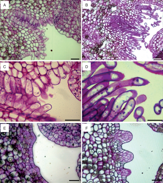
Fig. 5.
Changes in the ovary in ‘Nova’ mandarin. Comparison of the ovary at anthesis (A, C, E), and 10 d later (B, D, F). (A) Stylar canal lined with papillar cells come out into the ovary locule. (B) Papillae have elongated 10 d after anthesis and are approaching the ovule. (C) Short papillae at anthesis with starch grains. (D) Elongated papillar cells with secretion. (E) Ovary locule at anthesis with small protuberances. (F) Development of juice vesicles 10 d after anthesis. JB-4-embedded transverse sections of the ovary stained with PAS. Scale bars = 50 µm.

