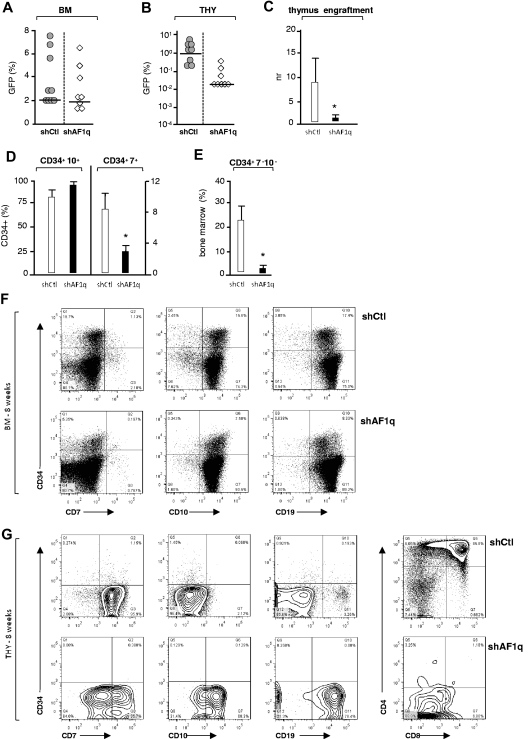
Figure 7.
Effect of AF1q deficiency on T-cell development in humanized mice. (A-B) NSG mice were injected with CD34+ HPCs (1.5 × 10⁵ cells) exposed to shAF1q or scrambled shRNA vectors and analyzed as described in Figure 6. The percentages of GFP+ cells among CD45+ human cells present in the BM (A) or thymi (B) of individual mice are shown (transduction efficiency: 12% ± 3%; 8 mice per condition, 2 experiments). Median percentages are indicated by a horizontal bar. (C) Effect of AF1q deficiency on thymus colonization. The efficiency of thymus engraftment was calculated and expressed as above. Statistically significant differences are marked by asterisks. (D-E) Percentages of CD7+ and CD10+ lymphoid precursors (D) or of multipotent CD7−CD10− HPCs (E) among CD34+GFP+CD45+ BM cells. Statistically significant differences are marked by asterisks. (F-G) Flow cytometric analysis of CD45+GFP+ cells in the BM (F) and thymi (G) of individual mice at week 8 after grafting. Numbers in quadrants indicate the percentages of each population.

