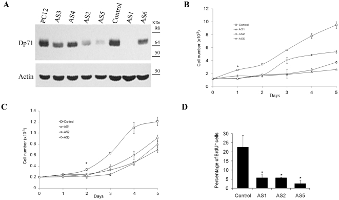
Figure 1. Knockdown of Dp71 expression decreases the proliferation of PC12 cells.
Dp71 protein levels were measured by western blotting in different Dp71-depleted clones (AS1–AS6) with the anti-Dp71 antibody 2166 and compared with those of PC12 cells and control cells (control). Membranes were stripped and reprobed with an anti-actin antibody for normalization. Migration of protein markers are denoted on the right (A). Cell proliferation of control and AS1, -2, and -5 cells was monitored over a 5-day period by both direct counting of viable cells via tripan blue exclusion (B) or MTT assay (C) and after 2 days of culturing, by BrdU incorporation using flow cytometry (D). Data are expressed as mean ± standard error of mean (SEM) of three independent experiments. Asterisks in Panel D denote significant differences (p<0.05).

