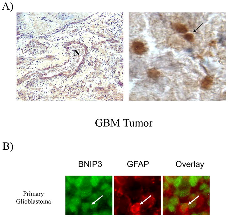
Figure 6. Malignant glial cells in GBM tumors express high levels of BNIP3 in necrotic regions that is primarily localized in the nucleus.
A) Primary glioblastoma multiforme (GBM) paraffin-embedded tumor tissues were immunostained by antibodies against BNIP3 (brown). DNA was counter-stained with hematoxylin (blue). The left panel shows a necrotic region (N) within a GBM immuno-stained for BNIP3 (brown) and counter-stained for DNA (blue). The arrow shows a cell expressing BNIP3. B) GBM tumor tissue was immunostained for BNIP3 (green) and GFAP (marker for glial cells, red). The slides were analyzed on a confocal microscope (Olympus). Arrows represent the same cell in each panel.

