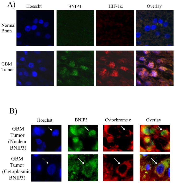
Figure 7. BNIP3 is expressed in hypoxic regions of GBM tumors but does not co-localize with the mitochondria.
A) Normal brain and primary GBM sections were immuno-stained with antibodies against BNIP3 and HIF-1α. DNA was stained with Hoescht dye and the slides were analyzed on a confocal microscope. These results represent three independent experiments. B) Glioblastoma tumors were immuno-stained with antibodies against BNIP3 (green) and cytochrome c (marker for mitochondria, red) as described in the Materials and Methods section. DNA was stained with Hoescht dye (blue). The images were captured on a fluorescent microscope with deconvolution software. Similar results were obtained from other GBM tumors. In each panel, arrows represent staining of the same cell expressing BNIP3.

