
Figure 1.
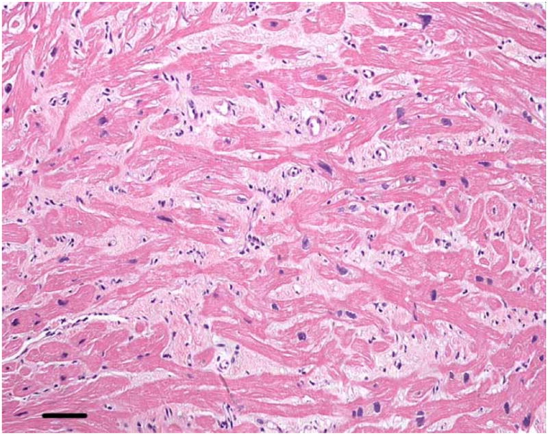
Right ventricular endomyocardial biopsy from Patient 1.
At high magnification the hypertrophied muscle fibers (dark pink) are separated by abundant interstitial fibrosis (light pink). Coursing in various directions, the myofibers in some areas are oriented orthogonally, characteristic of myofiber disarray. Hematoxylin and eosin, calibration bar = 45 microns. Color figure can be viewed in the online issue, which is available at www.interscience.wiley.com.