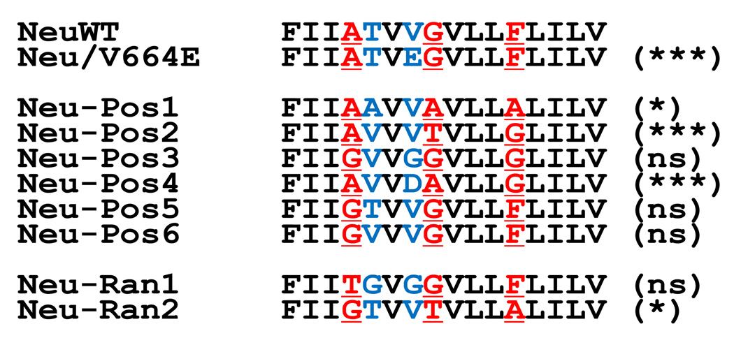
Figure 4. Sequences of Neu-related peptides.
Sequences of Neu/WT and Neu/V664E are shown at the top. Six dimeric sequences (Neu-Pos1 through Neu-Pos6) were identified in the screen. One of the seven sequences (Neu-Pos4) was identified twice independently. At the bottom are two random sequences from two beads out of the thousands of library members that were negative for dimerization in the screen (Neu-Ran1 and Neu-Ran2). In parenthesis is the statistical significance of the difference in activation in mammalian cells, relative to Neu/V664E, compared to the wild-type sequence, relative to Neu/V664E (ns=not significant, p> 0.05).

