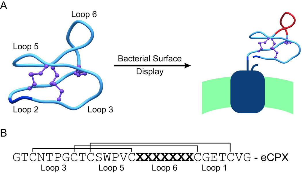
Figure 1.
Construction of a library of kalata B1 variants displayed on the cell surface of E. coli. A) The backbone of the cyclic kalata B1 peptide (PDB: 1NB1) was broken in loop two between two glycine residues (shown in dark blue) and fused to the display scaffold eCPX. Loop six was randomized to form the library (shown in red). B) Sequence of the kalata B1 peptide shown with the disulfide bond connectivity, the randomized region, and loop designations.

