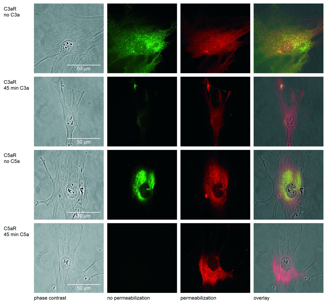
Figure 3.
Phase contrast and fluorescence images of immunostained C3aR and C5aR in osteoblasts without and after 45 min stimulation with 1µg/ml C3a or 0.1µg/ml C5a; first column: phase contrast images; second column: for the detection of membrane-bound receptors cells were fixed and the respective receptors were detected by a green fluorescent antibody as described in Materials and Methods; third column: for the detection of membrane-bound as well as internalized receptors cells were permeabilized before labelling with a red fluorescent antibody; fourth column: in the overlay the yellow colour indicates the membrane-bound receptors, whereas the internalized receptors are stained red.

