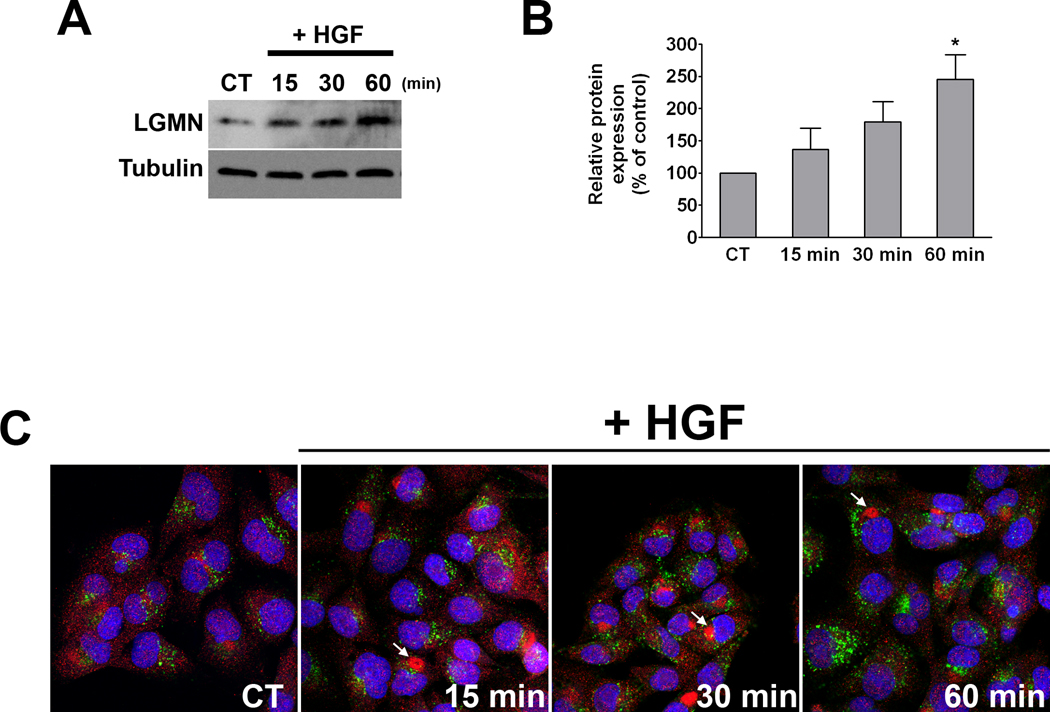
Figure 3. HGF stimulation increases LGMN expression.
(A) Western blot analysis of total cell lysates prepared from control (non-stimulated) cells and cells stimulated with HGF (100 ng/mL) for the indicated time periods demonstrates a time-dependent increase in LGMN expression. (B) Bar graph shows the densitometric quantification of 4 separate experiments (p<0.05, one-way ANOVA). (C) LGMN localization is altered after HGF treatment. SKHep1 cells were stimulated as above and examined by confocal immunofluorescence. LGMN (red) is present in punctate structures that do not co-localize with the lysosomal marker Lamp-1 (green), in either control or HGF-stimulated cells. Nuclei are labeled by TO-PRO-3 (blue). Upon HGF stimulation, LGMN accumulates near the nucleus (arrows).

