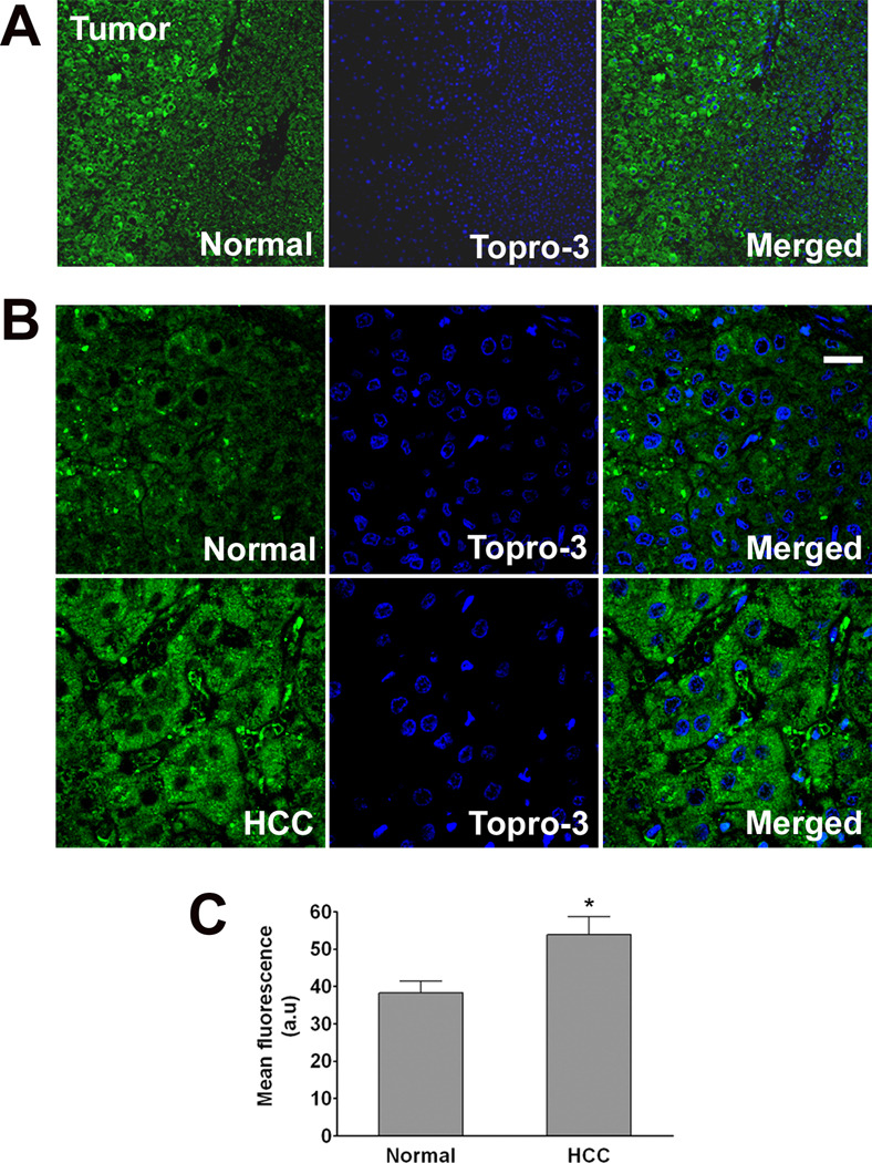
Figure 7. Expression of LGMN is increased in hepatocellular carcinoma (HCC).
Confocal immunofluorescence images were obtained from paraffin-embedded surgical specimens of tumors from patients with HCC. Immunohistochemical staining was performed to determine expression of LGMN (green) in tumor cells and in nearby normal hepatocytes. To-Pro-3 was used to identify cell nuclei (blue). (A) Low-power (10×) image of carcinoma cells and normal hepatocytes in the same field of view shows that LGMN staining is increased in the HCC. (B) Higher magnification (63×) images confirm increased expression of LGMN and show that it is distributed throughout the cytoplasm in HCC (Scale bar = 30 µm). Findings are representative of what was observed in 3 fields each of specimens from 5 separate patients. (C) Quantification of the average fluorescence in normal and HCC affected areas in the same specimen shows a significant increase in LGMN expression in the carcinoma cells (53.9±4.8 a.u.) as compared to normal hepatocytes (38.3±3.1 a.u.; p<0.01, paired t-test).

