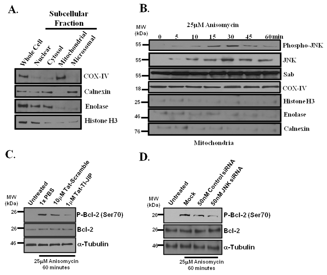
Figure 2. Anisomycin stress triggers JNK translocation to the outer mitochondrial membrane and JNK-mediated phosphorylation of Bcl-2.
(A) Representative mitochondrial enrichment and subcellular fractionation. Mitochondrial purity was determined by Western blotting for ER resident, calnexin; cytosolic enzyme, enolase; and nuclear protein, histone H3. Mitochondria were identified by COX-IV presence. (B) Anisomycin stress-induced JNK translocation to the mitochondria. Mitochondria were prepared from HeLa cells stressed with 25µM anisomycin for 0, 5, 10, 15, 30, 45, and 60 minutes. The mitochondria were monitored for Phospho-JNK, JNK, Sab, and COX-IV by Western analysis, and calnexin, enolase, and histone H3 were monitored as mitochondrial contaminants. COX-IV served as the mitochondria loading control. (C) HeLa cells were pretreated with either 10µM Tat-Scramble or 1µM TI-JIP for 30 minutes prior to incubation with 25µM anisomycin for 60 minutes. Cells were lysed and Western blot analysis was conducted for phosphorylation of Bcl-2 on serine 70, total Bcl-2, and loading control α-tubulin. (D) JNK was silenced with 50nM JNK-specific siRNAs or cells were treated with control siRNAs for 71 hours prior to addition of 25µM anisomycin for 60 minutes. Cells lysates were analyzed for the presence of Phospho-Bcl-2 (Ser70), total Bcl-2, or the loading control, α-tubulin.

