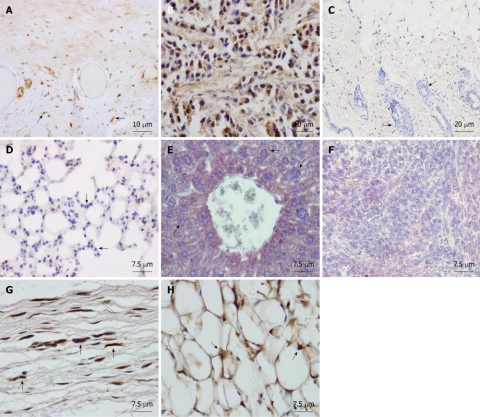
Figure 2.
Tissue sections from NOD/SCID mice transplanted subcutaneously with human adipose-derived mesenchymal stem cells. Mice were sacrificed at two mo and organs were formalin-fixed and paraffin-embedded. Sections were immunostained with monoclonal anti-human nuclear ribonucleoprotein antibody and counterstained with Harris hematoxylin. A: Human Positive control of the antibody, showing intense nuclear immunostaining in the testicular albuginea in brown. Arrows: human labeled fibroblasts; B: Positive control. SCID subepidermal teratoma showing positive nuclear staining for anti-human ribonucleoprotein. Arrows: human labeled teratoma cells; C: Saline injected SCID showing no staining for anti-human ribonucleoprotein in the skin at the site of injection. Arrows: mouse unlabelled cells of the hair follicles; D: Lung of an adipose-derived mesenchymal stem cell (AMSC) injected SCID mouse at 2 mo showing no human AMSC (hAMSC). Arrows: mouse unlabelled cells; E: Liver of an AMSC injected SCID mouse at 2 mo showing no hAMSC. Arrows: mouse unlabelled cells; F: Spleen of an AMSC injected SCID mouse at 2 mo showing no hAMSC. Arrows: mouse unlabelled lymphocytes; G: Subcutaneous connective tissue of an AMSC injected mouse at the site of injection, showing positive nuclear staining in the fibroblasts in brown (arrows); H: Subdermic adipose tissue of an AMSC injected mouse at the site of injection, showing positive nuclear staining in the adipocytes (arrows).

