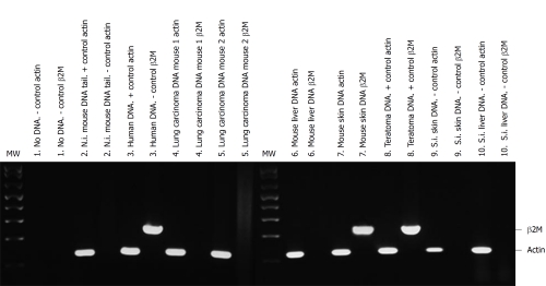
Figure 3.
Detection of human DNA from adipose-derived mesenchymal stem cells in NOD/SCID transplanted mice tissues. Human and mouse DNA were amplified by polymerase chain reaction with specific human β2 microglobulin primers (235 bp, β2M) and with actin primers that recognize mouse and human DNA (122 bp, actin) as mentioned in Materials and Methods. Each pair of lines shows the same DNA amplified with actin and with β2M primers. The following tissues of short-term transplanted mice (4 mo after cell transplantation) are shown: (1) Negative (-) control. No DNA in the polymerase chain reaction with actin or β2M primers; (2) Tail DNA from non-injected (N.i.) mouse tail DNA with actin (+ control of mouse DNA) or β2M primers (- control of human DNA); (3) Positive control. Human corneal tissue (human DNA) with actin or β2M primers; (4) Lung carcinoma DNA of a human adipose-derived mesenchymal stem cell (hAMSC) injected mouse 1 with actin or β2M primers; (5) Lung carcinoma DNA of another hAMSC injected mouse (2) with actin or β2M primers; (6) Liver DNA of a hAMSC injected mouse with actin or β2M primers; (7) Skin DNA of a hAMSC injected mouse with actin or β2M primers; (8) Teratoma DNA of NTERA-2 cells injected mouse with actin or β2M primers (positive (+) control for both sets of primers); (9) Negative control. Skin DNA from the saline injected (S.I.) mouse with actin or β2M primers; and (10) Negative control. Liver DNA from the saline injected mouse with actin or β2M primers.

