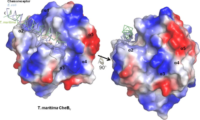
Figure 4. View of the proposed docking site with chemoreceptor α-helix shown over the T. maritima CheBc electrostatic potential surface.

View of the truncated α-helical substrate of the T. maritima and E. coli chemoreceptor overlaid on top of the T. maritima CheBc electrostatic potential surface are shown in two different orientations, which are rotated 90° with respect to each other along the indicated axis (Only T. maritima CheBc is shown for clarity). The α-helical substrate of the chemoreceptor sits within a groove created at the active site.
