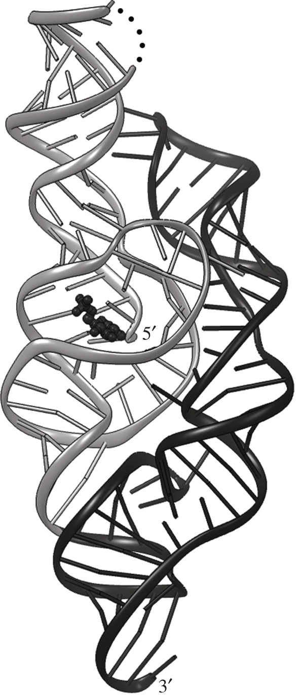
Figure 1.

Overall structure of the glmS ribozyme. Cartoon representation of the 1.7 Å resolution structure of the ribozyme from T. tengcongensis [6]. The doubly pseudoknotted core domain is coloured grey; the peripheral stabilizing domain black. The cofactor GlcN6P is shown as a black ball-and-stick model. Cleavage takes place between the 5′-most two nucleotides. Black dots depict a break in the RNA construct used for crystallization.
