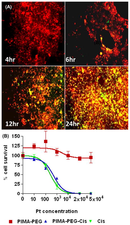
Figure 2.
In vitro characterization of PIMA-PEG-cisplatin nanoparticles. (A) Representative epifluorescence images of 4T1 breast cancer cells incubated with FITC-labeled nanoparticles for defined time periods. The cells were counterstained with Lysotracker red dye. Merge images reveal colocalization after 6 hours persisting up to 24 hours. (B) Concentration efficacy graph showing effect of empty polymer, cisplatin nanoparticle and free cisplatin on 4T1 cell viability at 48 h. Data shown are mean SEM from n=3 independent experiments with triplicates in each experiment.

