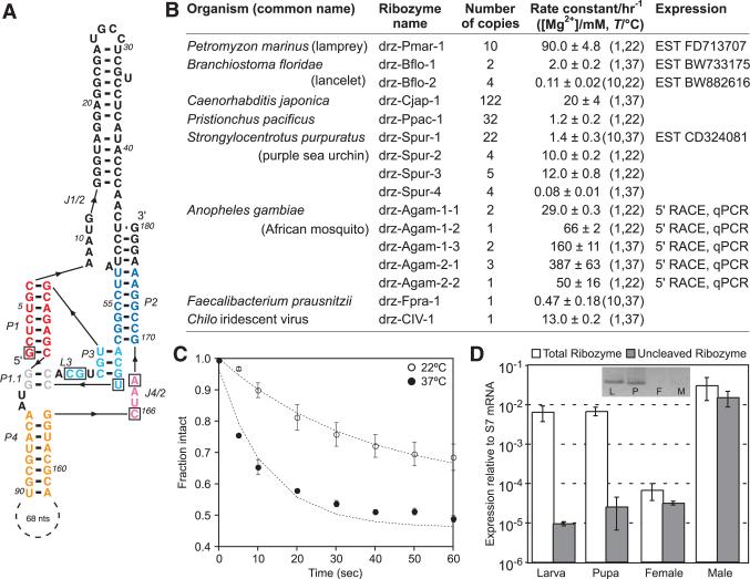
Fig. 1.
A. gambiae drz-Agam-2-1 ribozyme. (A) Core elements are colored by region corresponding to the HDV ribozyme (1). Boxed letters are the six invariant positions used in secondary structure searches that identified the ribozymes listed in (B). (C) Graph of in vitro self-cleavage in 1 mM MgCl2 and (D) RT-qPCR of drz-Agam-2-1 isolated from indicated developmental stages. (Inset) 5′ RACE of the ribozyme. All data are average values ± average deviations.

