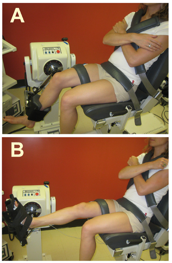
Figure 1.

Photographs of the experimental set-up for the assessment of the coactivation. Participants were comfortably secured in sitting position with the chest, pelvis and the tested leg being firmly anchored to the Biodex chair. A. For the knee extension task, the input axis of the dynamometer was adjusted to align with the knee axis of rotation through the lateral femoral condyle and the foot was left free. B. For the plantarflexion task, the ankle joint (lateral malleolus) was aligned with the input axis of the dynamometer and fixed in plantarflexion (110°) using the standard Biodex ankle unit attachment. EMG activity was recorded from vastus lateralis (VL), rectus femoris (RF), soleus (Sol) and gastrocnemius lateralis (GL).
