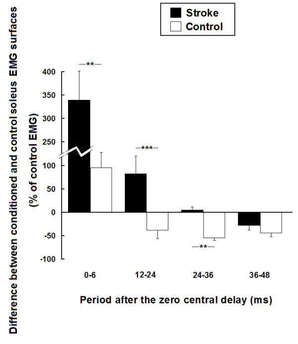
Figure 6.

Group comparisons of the effects of femoral nerve stimulation on soleus voluntary EMG activity in 13 stroke and 10 healthy participants. Mean modulations of soleus voluntary EMG activity induced by femoral nerve stimulation for the stroke group (black bars) and the control group (white bars) (expressed as a % of soleus unconditioned EMG surface). Modulations are presented within the four time windows of analysis from 0 to 6 ms, 12 to 24 ms, 24 to 36 ms and 36 to 48 ms after the zero central delay. Positive values (i.e. above zero on the ordinate) denote facilitation and negative values denote inhibition. Vertical bars = 1 SEM. Asterisks represent significant difference in modulation between the control and the stroke participants (* p ≤ 0.05; ** p ≤ 0.01; *** p ≤ 0.001).
