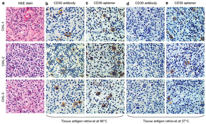
Figure 2.

Tissue immunostaining of classical Hodgkin lymphoma (CHL) by the CD30 aptamer probe and the CD30 antibody. CHL tumor cases were selected with hematoxylin and eosin stain for morphological confirmation (a). Tissue antigen retrieval was performed at 96°C, and lymphoma cells were probed with CD30 antibody for 90 min or CD30 aptamer probe for 20 min (b and c). The same sets of tissue immunostains were also performed with antigen retrieval at 37°C (d and e). All images were taken under a light microscope with ×200 magnification.
