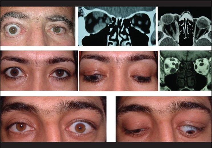
Figure 1.

Top photos show a patient with unilateral thyroid optic neuropathy and coronal and axial computerized tomography (CT) scan demonstrating an apical crowding. Middle photos show a patient with unilateral thyroid extra-ocular myopathy and coronal CT scan demonstrating muscles’ enlargement. Bottom photos show a patient with unilateral thyroid eyelid retraction
