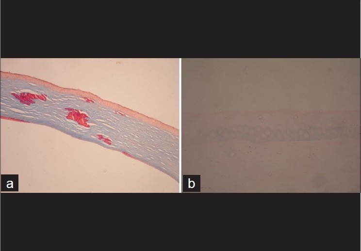
Figure 4.

(a) Section of the cornea shows bright-red crystalline deposits in the stroma characteristic of granular dystrophy. (b) The basal cells show intracytoplasmic Prussian blue reaction with Perl's stain, confirming the presence of iron deposits, a characteristic feature of keratoconus
