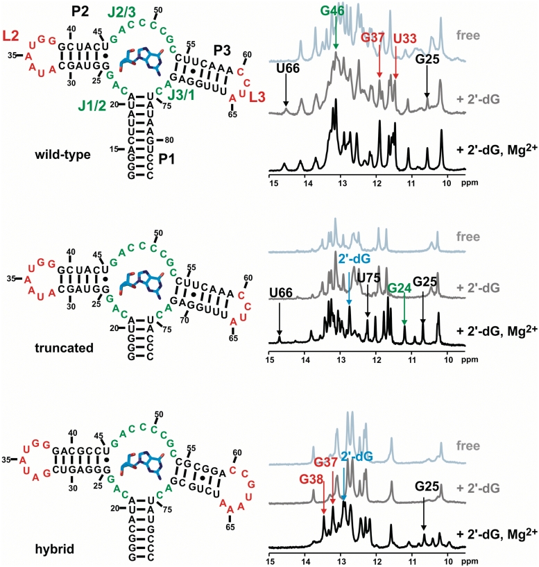
Figure 1.
Secondary structures of the three RNA constructs wild-type (upper left), truncated (middle left) and hybrid (lower left) aptamer. On the right side, the corresponding 1D-NMR titration spectra are shown (light grey: free aptamer, dark grey: 2′-dG added, black: 2′-dG and 5 mM Mg2+ added). Arrows indicate key resonances as reporters of ligand-binding induced folding. Nucleotide numbering is according to the purine aptamer nomenclature originally proposed (33).

