Figure 2.
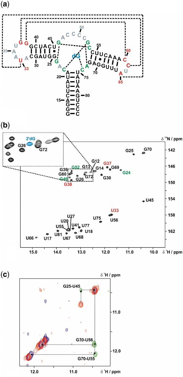
NMR characterization of the mfl-aptamer-2′-deoxyguanosine complex. (a) Secondary structure of the wild-type mfl-aptamer with the native ligand 2′-dG, resonances in non-helical regions detectable by NMR are colour-coded (red: loop regions; green: binding pocket region). Long-range interactions are indicated by dashed lines. (b) 1H,15N-HSQC of the mfl-aptamer in complex with 2′-dG. Signal annotations are colour-coded corresponding to the region of their assigned residues. The small section of the guanosine imino proton region of a 15N- HSQC with 15N-labelled ligand 2′-dG shows the ligand imino proton resonance in blue. (c) Spectral region of the double-half-filtered NOESY showing an overlay of the 14N(ω1, ω2)-edited signals in blue, the 15N(ω1, ω2)-edited signals in red and the 14N(ω1),15N(ω2)-edited signals in green. With a 15N-guanosine-labelled sample, ambiguities arising from spectral overlap were resolved.

