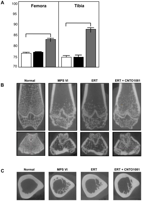
Figure 3. Bone length and microarchitecture in untreated and treated MPS VI rats.
(A) MPS VI rats were subjected to the ERT (black) or combined ERT/anti-TNF-alpha (gray) treatment for 8 months, as described in the text (n = 8/group). The animals were euthanized 2 days after the last injection, and the femora and tibia were collected for microCT analysis. The results were compared to untreated age and gender-matched MPS VI rats (white), and the values expressed as a percentage of normal controls. ERT did not increase the length of the femora or tibia, while the combined protocol led to increases of ∼6 and 14%, respectively. Of note, the tibia and femora in the combined treatment group were on average ∼88 and 84% of normal, compared to 74 and 77% in the untreated MPS VI group. (B) microCT analysis of the coronal views. In untreated and treated MPS VI rats the trabecular density within the metaphyseal bone was reduced, the physeal growth plate was dysmorphic and disrupted, and the epiphyseal trabeculae were disorganized relative to the normal femora. Mild improvement from the combined treatment was detected. (C) microCT analysis of the mid-diaphyseal region of the femora showing axial views with subcortical trabecular infiltration into the marrow space. Although some reductions in trabecular infiltration were noted following treatment, these could not be confirmed by quantitative measures.

