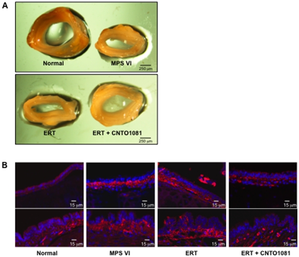
Figure 4. Tracheal defects in untreated and treated MPS VI rats.
(A) Tracheas were collected from treated and untreated MPS VI and normal animals at the end of the study (37 weeks of age). As illustrated by this representative figure, untreated MPS VI rats had markedly thickened and abnormal, collapsed tracheas with narrow, flattened interior openings. These abnormalities were not altered by ERT, but were clearly improved by the combined treatment which resulted in rounded tracheas with almost statistically normalized cross sectional areas. (B) Immunohistochemical analysis of the tracheas showed increased expression of the pro-inflammatory and pro-apoptotic sphingolipid, ceramide, in the epithelial cells of untreated and ERT-treated animals (red), consistent with the occurrence of inflammatory disease. Tracheas from the combined treatment group showed almost normal ceramide expression.

