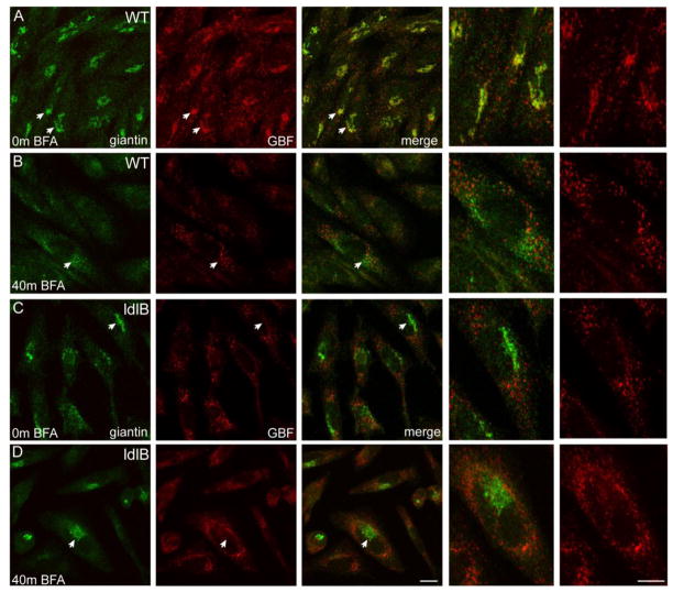
Figure 2. ldlB CHO cells are resistant to the Golgi-disrupting effects of BFA.
Wild type and COG1-deficient ldlB CHO cells were cultured in the absence (A,C) or presence (B,D) of 5 μg/mL for 40 min, co-stained with giantin and GBF1 antibodies and analyzed by confocal microscopy. Although GBF1 is readily detected on the Golgi in WT cells (A), very little Golgi staining of GBF1 was observed in the ldlB cells (C). Collapse of the Golgi marker giantin is observed in WT cells following BFA treatment (B) but the ldlB cells are resistant (D). Arrowheads denote cells that are enlarged in the far right panels. Bar = 10 μm.

