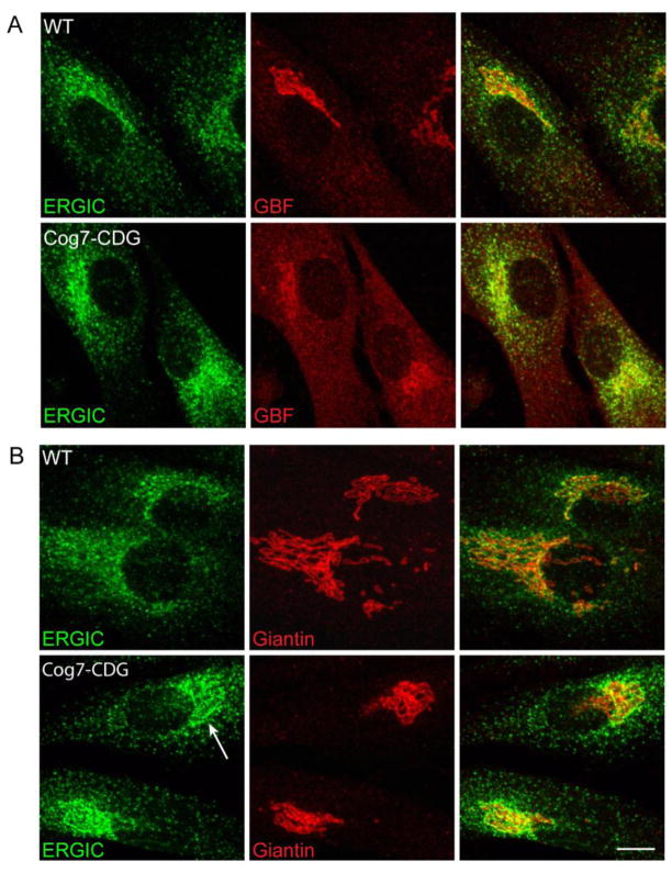
Figure 5. The redistribution of GBF1 to the ERGIC in Cog7-deficient fibroblasts may be masked by the juxtaposition of ERGIC and Golgi membranes.
WT and Cog7-deficient fibroblasts were co-stained with antibodies against ERGIC-53 and GBF1 (panel A) and giantin (panel B). Compared to wild type cells, more peripheral GBF1 staining is observed in the Cog7-deficient fibroblasts as well as a shift of the GBF1-positive ERGIC membranes closer to the cis-Golgi face. Bar = 10 μm.

