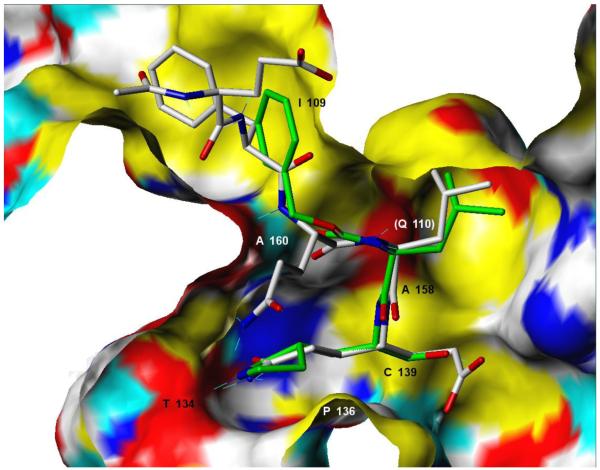
Figure 3.
Predicted covalently-bound conformer21 for NV 3C protease inhibitor 4 (stick structure with green carbon atoms and CPK-colored N and O atoms) contrasted with the peptidic inhibitor acetyl-Glu-Phe-Gln-Leu-Gln-CH=CH-COO− (stick structure with gray carbon atoms and CPK-colored N and O atoms) resolved in the 1IPH crystal structure.7 The NV 3C protease binding site is shown as a Connolly surface colored as follows: yellow = non-polar groups, white = partially polar C, H atoms, red = polar O, blue = polar N, cyan = polar H. Key pharmacophore residues are labeled according to the positions on the receptor surface from which they interact with the ligand (except for Q110 whose approximate position is marked but whose surface is not shown because the residue is above the plane of the molecule).

