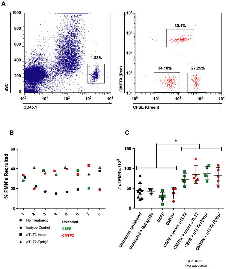
Figure 6. TLT2 ligation enhances neutrophil recruitment into sites of inflammation in vivo.
(A) Purified neutrophils from CD45.1 mice were labeled with either CFSE, CMPTX, or left unlabeled. These cells were then incubated in the presence of the αTLT2 mAb as indicated, or an isotype control antibody, or medium alone. After treatment, the purified, labeled neutrophils were mixed at equivalent ratios and 3×10⁷ total cells were adoptively transferred into CD45.2 recipient mice, which had received an intratracheal challenge with 5 μg of LPS to induce inflammation in the lung 30 min prior to adoptive transfer. After 4 h, the recipient mice were sacrificed and the neutrophils present in the lung were isolated by lavage. Donor derived neutrophils were indentified based on the expression of CD45.1, and these cells were analyzed for the presence of the fluorescent dyes, which discriminated the pre-transfer treatment conditions. (B) Ligation of TLT2 increases the relative frequency of neutrophils recruited into the lungs of recipient mice. A total of 8 mice are depicted and for each mouse the % PMNs recruited to the lung for each of 3 pre-treatment conditions is shown. The pretreatment condition is coded by the symbol used and the dye used is reflected by the color of the symbol. (C) TLT2 ligation causes an absolute increase in the number of neutrophils that migrate into inflamed lungs. The absolute number of neutrophils for each treatment condition was calculated based on flow cytometric analysis. The mean ± SD for a minimum of 5 mice is shown for each treatment condition.

