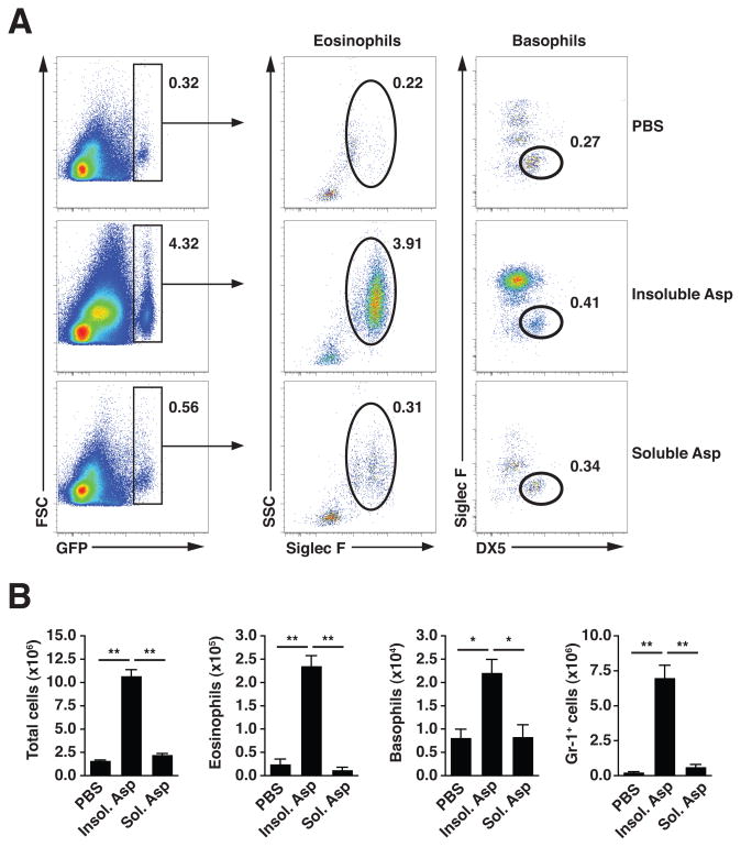
Figure 2. Intranasal challenge with insoluble components of fungal preparation induces accumulation of innate effector cells.
(A) Flow cytometric analysis of GFP-expressing lung eosinophils (GFP+SiglecF+SSC+) and basophils (GFP+SiglecF−DX5+) in 4get mouse lung tissue 1 day after 2 intranasal challenges with soluble or insoluble fractions from A. niger preparation (Asp). Numbers indicate the percentage of total live cells (DAPI−, not shown), and are representative of 3 independent experiments. (B) Total live cell numbers, with eosinophil and basophil subsets calculated from flow cytometry percentages as shown in (A), or percentage Gr-1+ cells (not shown). Mean±SEM, n=3/group; *p<0.01, **p<0.001, unpaired t-test.

