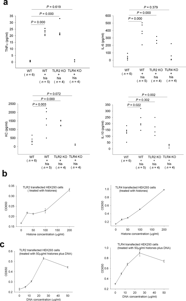
Fig.1. Extracellular histones trigger TLR2 and TLR4 signaling.
(a) TNFα, IL-6, KC and IL-10 levels in wild type, TLR2 KO or TLR4 KO mouse plasma collected 2 h after an intravenous injection of calf thymus histones (25 mg per kg body weight). Data from 5 experiments. (b) Reporter gene expression levels in TLR2 or TLR4 transfected HEK293 cells stimulated with calf thymus histones at the indicated concentrations. The results shown are the means ± SD performed in triplicate. (c) Reporter gene expression levels in TLR2 or TLR4 transfected HEK293 cells stimulated with calf thymus histones (50 µg/ml) and calf thymus DNA at the indicated concentrations. The results shown are the means ± SD performed in triplicate.

