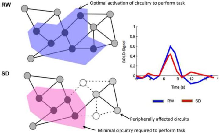
Fig. 6. Schematic illustrating how ‘off’ state neurons in awake individuals may affect performance during SD.
Each node represents a neuron and each solid line edge, a functional connection. During RW, ‘neurons’ in the area shaded blue, are activated. They represent the optimal level of activation of neural circuits required to perform the task. During SD, ‘neurons’ in this network represented in open circles go into an ‘off’ state, leaving only a critical minimal circuitry, to execute the task (area shaded pink). This could lead to less efficient processing, or if more neurons enter the ‘off’ state, a brief inability to respond. The light blue ‘neurons’ indicate how other neurons not immediately affected during task performance may be peripherally affected by the ‘off’ neurons going offline.

