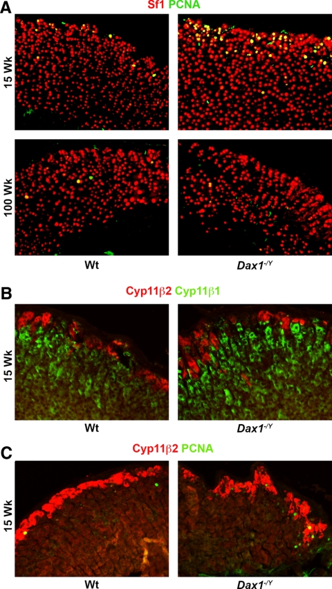
Fig. 6.
Visualization of PCNA reactivity and cortical zonation in young and old Wt and Dax1−/Y mice. Adrenal sections from mice of the indicated ages were subjected to immunofluorescence as described in Materials and Methods using (A) antibodies directed against PCNA (green) and Sf1 (red) and (B) antibodies directed against Cyp11β2 (red) and either cyp11β1 or PCNA (green). Images were merged to show colocalization.

