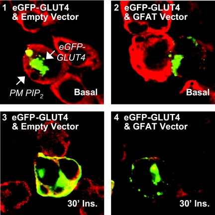
Fig. 6.
Overexpression of GFAT reduces detectable cell-surface PIP2 concomitant with a reduction in insulin-stimulated GLUT4-eGFP translocation. Differentiated 3T3-L1 adipocytes were electroporated with 50 μg GLUT4-eGFP cDNA and 200 μg GFAT cDNA empty vector or GFAT cDNA. The cells were allowed to recover for 16 h. Cells were subsequently incubated in serum-free medium for 2 h and then left untreated (basal, panels 1 and 2) or treated (30′ Ins., panels 3 and 4) with 100 nm insulin for 30 min. Cells were fixed, labeled for PIP2, and subjected to confocal fluorescence microscopy. Representative images from three independent experiments are shown.

