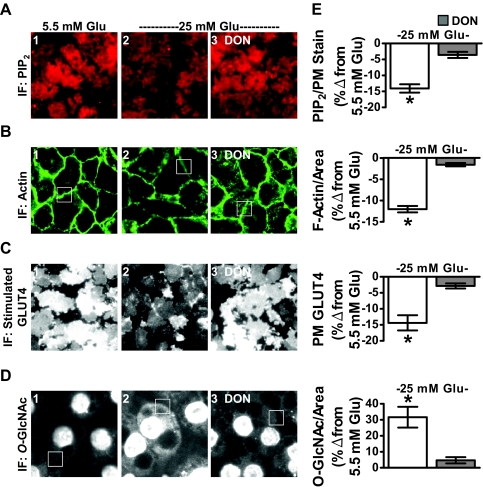
Fig. 7.
High glucose alone induces PIP2/F-actin loss and insulin resistance in 3T3-L1 adipocytes that were cultured and differentiated in 5.5 mm glucose. Murine 3T3-L1 preadipocytes cultured and differentiated in DMEM containing 5.5 mm glucose were left untreated (5.5 mm Glu, panel 1) or treated overnight (16 h) with 25 mm glucose (25 mm Glu, panels 2 and 3) in the absence or presence of DON (panel 3). PM PIP2 (A), F-actin (B), insulin-stimulated PM GLUT4 (C), and O-linked glycosylation (D) were determined and quantitated (E) as described in preceding figures. All microscope and camera settings were identical between groups, and representative images from three independent experiments are shown. Values are means ± se from three independent experiments. IF, Immunofluorescence. *, P < 0.05 vs. control).

