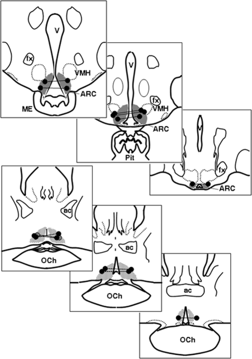
Fig. 2.
Sites of microimplants in study 1. The top three panels depict bilateral microimplants in the ARC; the bottom three panels are sites in the POA. Bilateral sites in the same ewe are connected by a line. The shaded area depicts distribution of PR-containing dynorphin neurons based on previous data (25). Ac, Anterior commissure; fx, fornix; ME, median eminence; OCh, optic chiasm; Pit, pituitary stalk; VMH, ventromedial hypothalamus.

