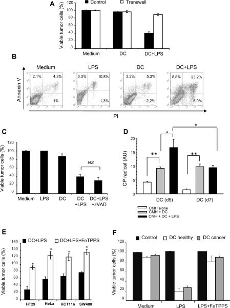
Figure 3. Human monocyte-derived DC killing activity depends on peroxynitrites.
A: HT29 cells were cultured alone (Medium) or with monocyte-derived DC from healthy donors, in the presence (DC+LPS) or not of LPS (DC). The cells were cultured together (Control) or separated by a transwell insert (Transwell). Tumor cell killing was determined after 48 h. B: HT29 tumor cells were cultured alone (Medium), with LPS (LPS), with day 5 monocyte-derived DC from healthy donors with (DC+LPS) or without LPS (DC). After 48 h, the cells were stained with CD1a Ab and with annexine V-FITC/propidium iodide (PI). The percentage of PI−/Annexine-V+ (apoptotic) or PI+/Annexine-V+ (necrotic) tumor cells was determined after gating on CD1a negative HT29 tumor cells. C: HT29 tumor cells were cultured alone (Medium), with LPS (100 ng/ml) (LPS), with day 5 monocyte-derived DC (DC), with DC in the presence of LPS (DC+LPS) or DC in the presence of LPS + z-VAD-fmk (40 μM) (ratio DC:tumor cell = 5:1). Tumor cell survival was determined after 48 h. D: Production of reactive oxygen species by non activated or LPS-activated human monocyte-derived DC was determined by resonance electron spin. CMH was used as the spin probe. Day 5 monocyte-derived DC from healthy donors were cultured with (DC+LPS) or without LPS (DC) for 6 h. Activated and non-activated human DC (2×10⁶) were harvested and incubated with 1 mmol/l CMH. The amplitude of center field anisotropic signal was quantified in arbitrary units in order to determine CP• nitroxide formation in LPS-activated or non-activated human dendritic cells at day5 and day7. This experiment was done 3 times with DCs from 3 different healthy donors and from 3 cancer patients.*, p<0.05; **, p<0.005. E: HT29 tumor cells were cultured alone (Control) or with day 5 monocyte-derived DC from healthy donors (DC): without LPS (Medium), with LPS (LPS) or with LPS and FeTPPS (50μM) (LPS+FeTPPS). Tumor cell viability was determined after 48 h. F: Different human tumor cell lines (HT29, HeLa, HCT116, SW480) were co-cultured with LPS-activated human monocyte-derived DC from healthy volunteers or from cancer patients in the presence (DC+LPS+FeTPPS) or absence (DC+LPS) of FeTPPS (50μM) (DC:tumor cell ratio = 5:1). Tumor cell viability was determined after 48 h. Similar results were obtained with DC generated from 6 healthy donors and 4 cancer patients. *, Significant difference when compared with tumor cells cultured with activated DC in the absence of FeTPPS (p<0.05).

