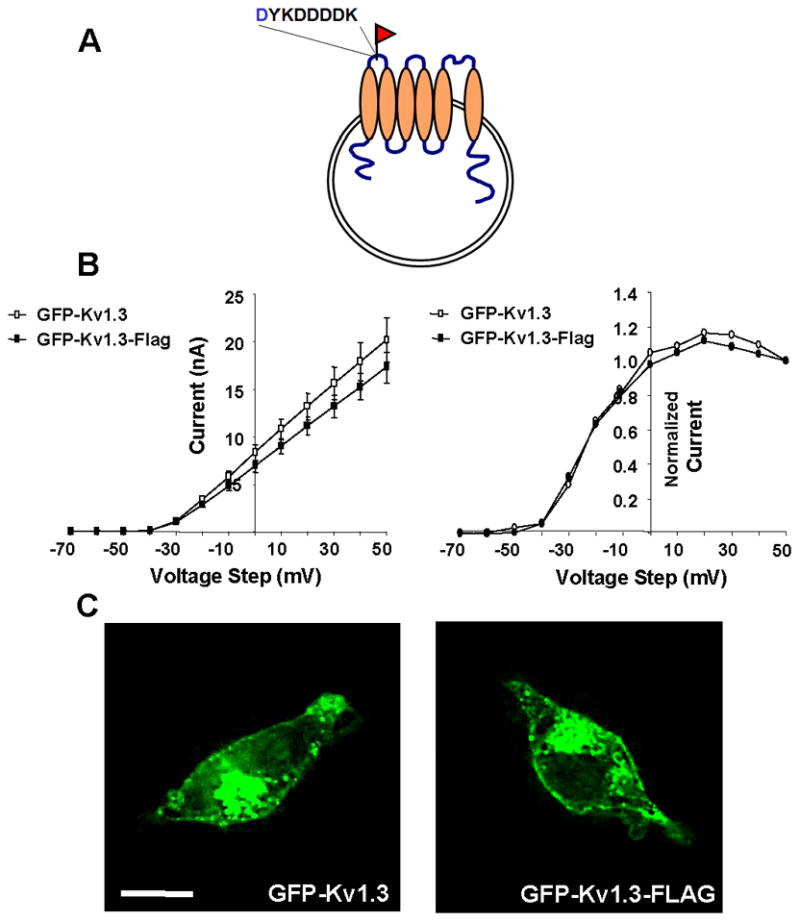
Fig. 1. Incorporation of extracellular FLAG epitope into GFP-Kv1.3 for studying Kv1.3 localization.

A FLAG epitope was inserted into an extracellular region of wild type and mutant GFP-Kv1.3 to allow detection of Kv1.3 at the cell surface. A) Schematic showing where the FLAG epitope was inserted. B) Steady-state current-voltage relationships for GFP-Kv1.3 (open symbols) and GFP-Kv1.3-FLAG (closed symbols) are shown on left and indicate no effect of the FLAG epitope (n = 8; p > 0.05; unpaired t-test). Activation curves plotted from the tail current of GFP-Kv1.3 (open symbols) and GFP-Kv1.3-FLAG (closed symbols) are shown on the right and indicate that the FLAG epitope did not affect voltage sensitivity (n = 8; p > 0.05; unpaired t-test). C) Fluorescence micrographs of HEK293 cells transfected with GFP-Kv1.3 and GFP-Kv1.3 FLAG. Scale bar represents 10 μM.
