
Figure 2. Comparison of Nissl, somatostatin, and parvalbumin staining in CA1.
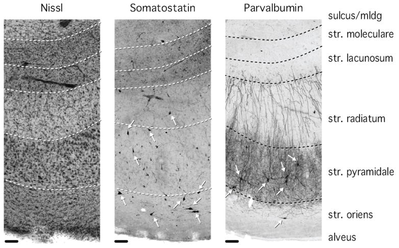
Cell bodies stained for somatostatin are located predominantly in stratum pyramidale and stratum oriens, with occasional appearance of cell bodies in stratum radiatum. Somatostatin processes are particularly dense in stratum lacunosum/moleculare. Cell bodies stained for parvalbumin are located predominantly in stratum pyramidale and to a lesser extent in stratum oriens. Parvalbumin-positive processes are densely distributed in stratum pyramidale and less densely in stratum radiatum. Arrows point to stained neuronal cell bodies. Str = stratum, mldg = molecular layer of the dentate gyrus. Scale bar: 100 μm.