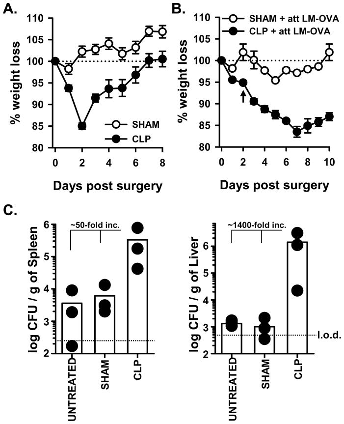
Figure 1.
CLP-treated WT B6 mice demonstrate increased morbidity and susceptibility to LM-OVA infection. A. WT B6 mice underwent sham or CLP surgery. Animal weight was measured daily beginning the day of surgery, and the percent weight loss with respect to the starting weight is shown. B. Sham or CLP surgery was performed on WT B6 mice. On d 2 post-surgery, the mice were infected with 107 CFU attenuated LM-OVA. Animal weight was measured daily beginning the day of surgery, and the percent weight loss with respect to the starting weight is shown. In A. and B., the dotted line represents the starting weight of the mice, normalized to 100%. In B., the arrow indicates the time of infection. C. Sham or CLP surgery was performed on WT B6 mice, and the mice were infected with attenuated LM-OVA on d 2 post-surgery as in B. In addition, naïve mice that did not undergo any surgery (“untreated”) were also infected with attenuated LM-OVA infection. The bacterial titer in the spleen and liver were then determined on d 3 post-infection. The dotted line represents the limit of detection (l.o.d.).

