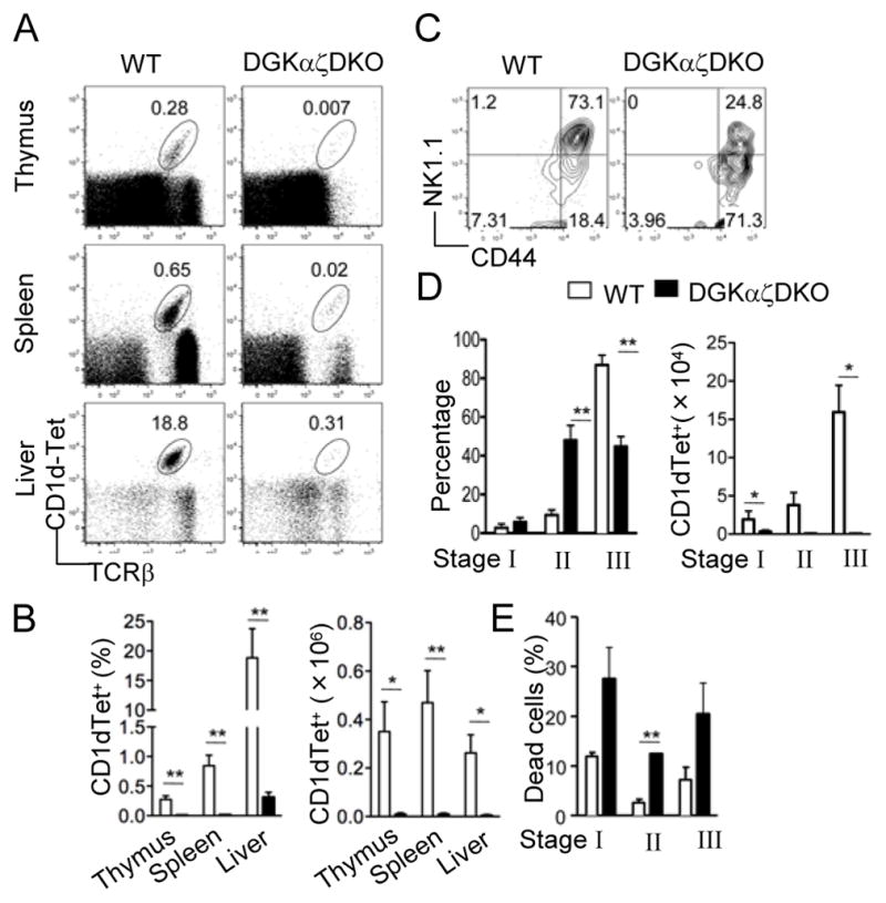
Figure 2.

Severe iNKT cell developmental defects in DGKαζDKO mice Thymocytes, splenocytes, and liver mononuclear cells from age- and sex-matched DGKαζDKO mice and WT controls were subjected to flow cytometric analysis. Data shown are representative of five mice per group. (A) Flow cytometry of cells stained with CD1d-Tet and anti-TCRβ. (B) Percentage (left) and number (right) of live CD1d-Tet+ TCRβ+ cells (mean, s.e.m.). (C) Expression of CD44 vs. NK1.1 on live CD1d-Tet+CD24− gated thymocytes. (D) Percentage (left) and number (right) of CD1d-Tet+CD24− live thymocytes in different iNKT developmental compartments (mean, s.e.m.). (E) Percentage of cell death (defined by positive Live-Dead® staining) in different iNKT developmental compartments (mean, s.e.m.). *, p<0.05; **, p<0.01; ***, p<0.001.
