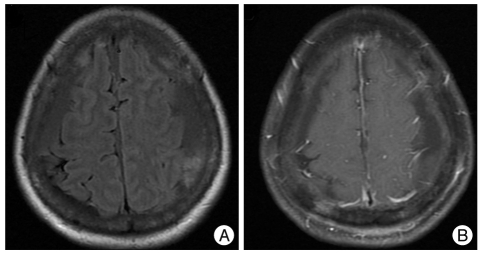
Fig. 2.
Magnetic resonance (MR) imaging 1.5 month after the seizure attack. A : A bilateral multi-staged subdural hemorrhage is detected, whereas the lesion in the parieto-occipital lobe is no longer present in the T2-weighted and FLAIR axial MR imaging. B : There is no enhancement in the gadolinium enhanced T1-weighted axial MR imaging. FLAIR : fluid attenuated inversion recovery.

