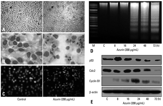
Fig. 3.
Azurin triggers apoptosis of YD-9 cells. Morphological changes in YD-9 cells after 24 h incubation in the presence or absence of 200 µg/mL azurin, as observed with simple microscopy (A) and after cell staining with hemacolor (B) or Hoechst 33352 (C). (D) DNA fragmentation of 200 µg/mL azurin-treated cells occurred in a time-dependent manner. (E) YD-9 cells were exposed to 200 µg/mL azurin for indicated times. Total protein (30 µg) was subjected to SDS-PAGE and western blotting. Data shown is the representative of 3 independent experiments. M, DNA marker; C, Control (lysate of untreated cells).

